
KNEE3D
KNEE3D
At KNEE3D our mission is to modernise imaging of the knee joint away from 2D X-rays using state-of-the-art 3D analysis so that everyone can see and understand knee joint health better.
We believe that 3D computed tomography (CT) of the knee is the future, holding valuable, untapped information that has superior detail and reliability compared to X-rays when assessing joint diseases such as osteoarthritis.
Starting with the provision of research analysis services and expert consultancy, our plan is to grow and provide point-of-care 3D evaluation of CT imaging that will replace 2D X-rays, helping everyone to make the best possible choices for their knee joint health.
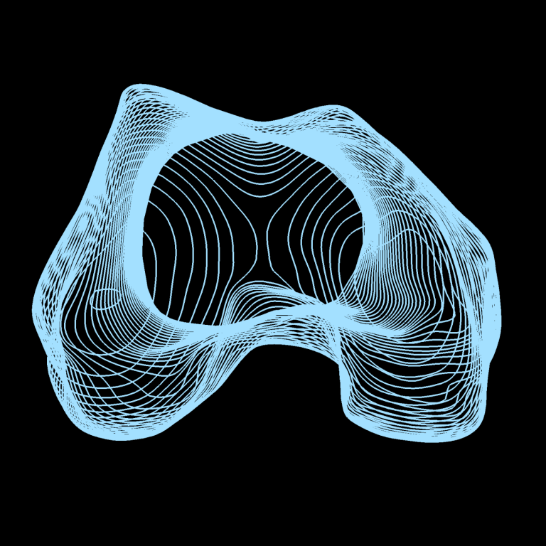
We provide reliable and robust 3D visualisation and analysis of the knee joint from CT imaging data using state-of-the-art techniques.

We see partnership as essential for developing ideas with the greatest impact. Join us to shape the future of knee joint imaging with CT.

Our goal is for KNEE3D to help everyone concerned about knee joint health make the best possible lifestyle and treatment choices.
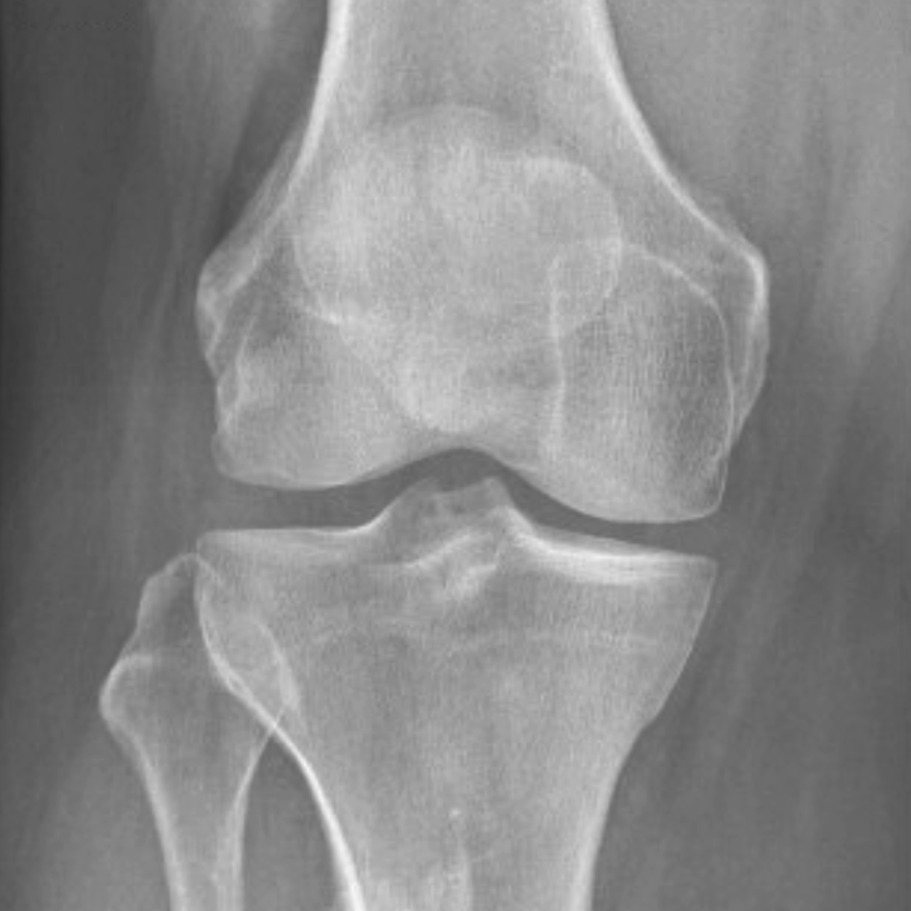
Frontline imaging of the knee is usually with X-ray, a flattened 2D image of the bones and joint space. Our mission is to replace 2D X-ray imaging with 3D CT as standard. Image credit Vecteezy
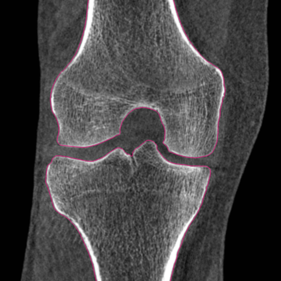
From a simple scan taking a few seconds, CT delivers a 3D data volume that can be viewed in every direction. Here we see just one of many possible slices through the knee with bones outlined in pink from our segmentation.
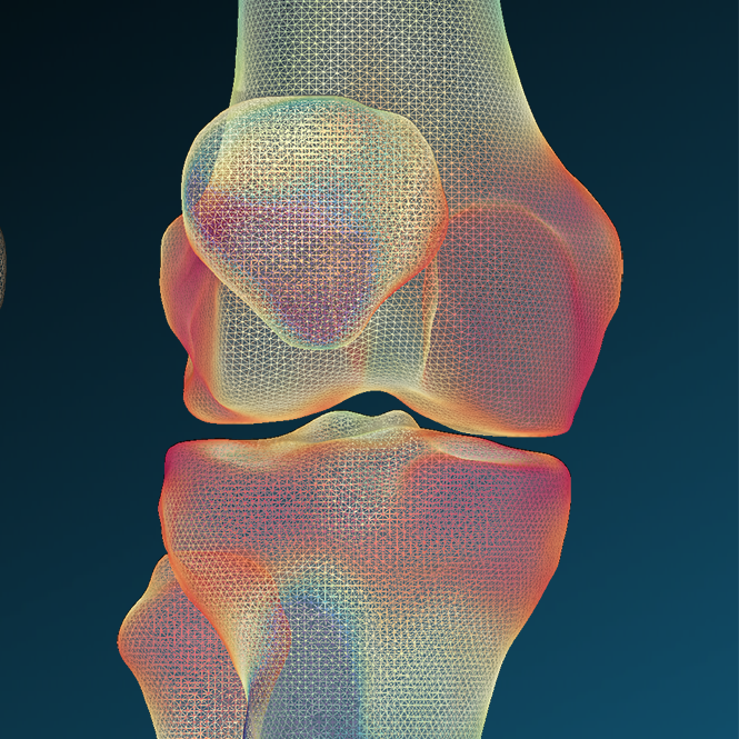
Using CT imaging data, we can build 3D models of the knee joint, accurately and reliably measure different bone properties, and then map these across the bone surfaces in a way that can't be done with X-ray imaging.
We need your consent to load the translations
We use a third-party service to translate the website content that may collect data about your activity. Please review the details in the privacy policy and accept the service to view the translations.